Small intestine
Organ where most nutrient absorption occurs in digestion.

BruceBlaus . When using this image in external sources it can be cited as: Blaus · CC BY 3.0
The small intestine, also known as the small bowel, is an organ in the gastrointestinal tract where most of the absorption of nutrients from food takes place. It lies between the stomach and large intestine, and receives bile and pancreatic juice through the hepatopancreatic ampulla and major duodenal papilla to aid in digestion. The small intestine is about 6.5 metres (21 feet) long in post-mortem measurements, but in a living person its length typically ranges from 3 to 5 metres (10 to 16+1⁄2 feet), folding many times to fit in the abdomen. Although it is longer than the large intestine, it is called the small intestine because it is narrower in diameter.
- length_typical_living_person
- 3–5 m (10–16+1⁄2 ft)
- diameter_adult
- 2.5–3 cm (1–1+1⁄8 in)
- parts
- duodenum, jejunum, ileum
- blood_supply
- celiac trunk and superior mesenteric artery
- key_function
- absorption of nutrients
Lore & Background
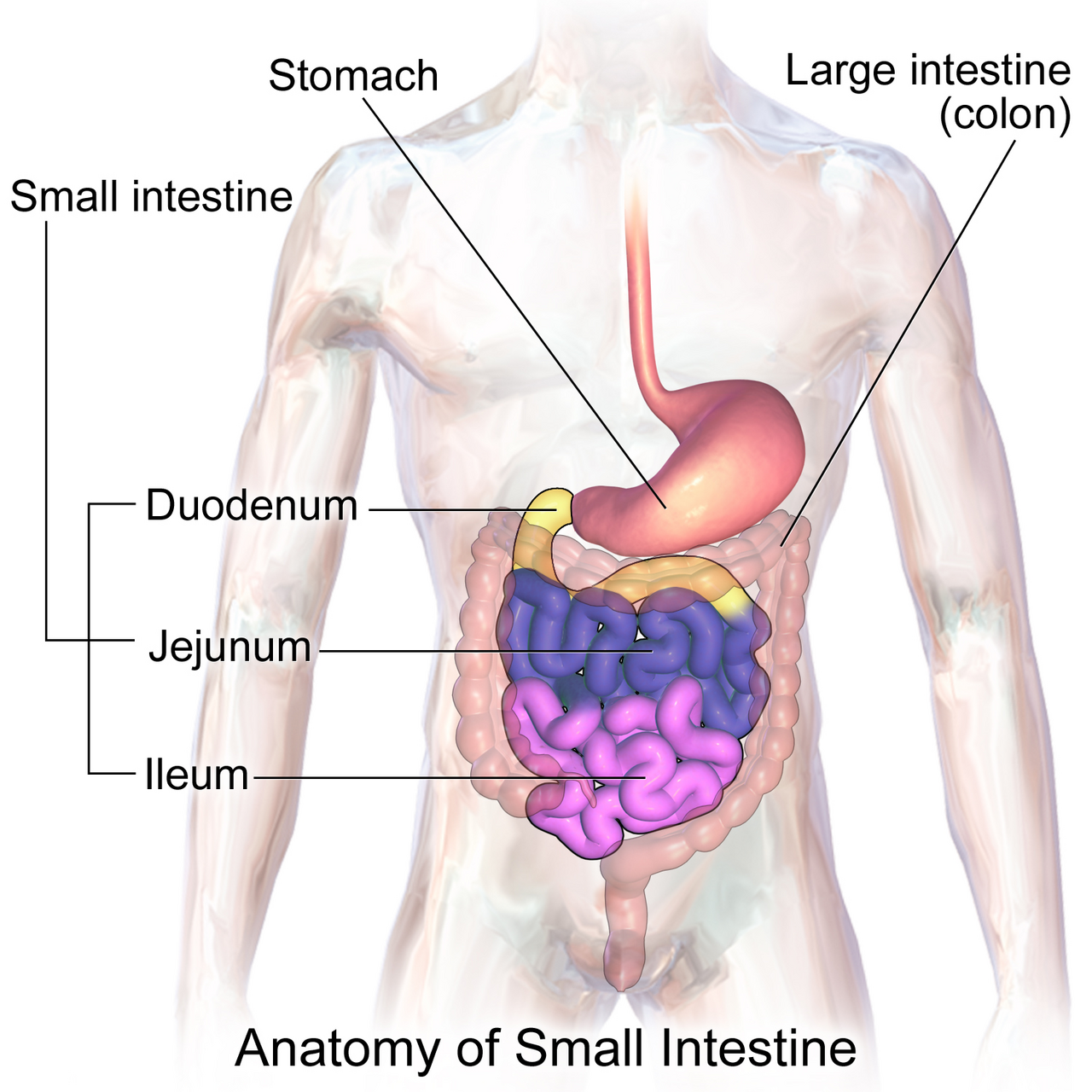
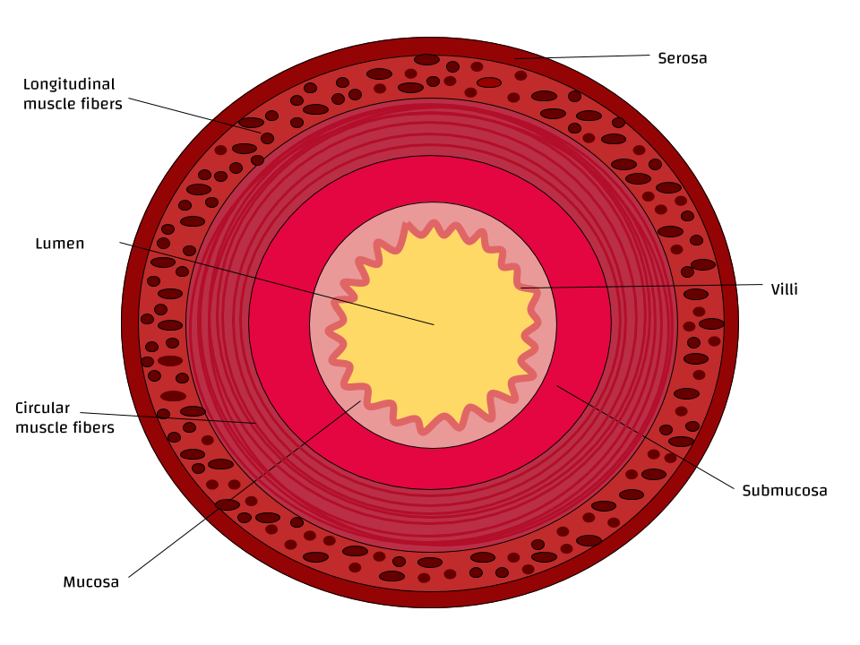
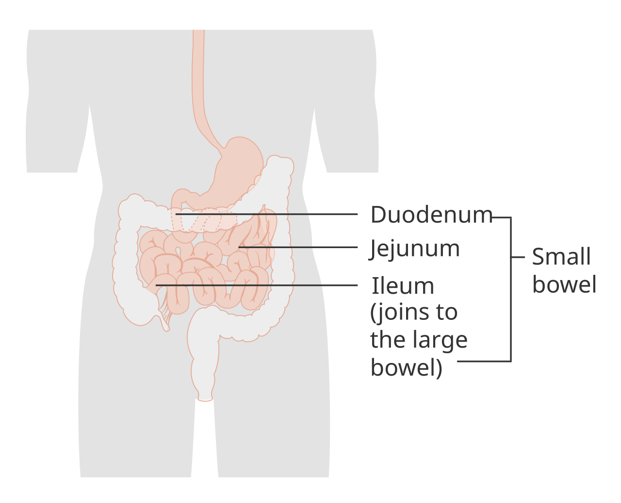
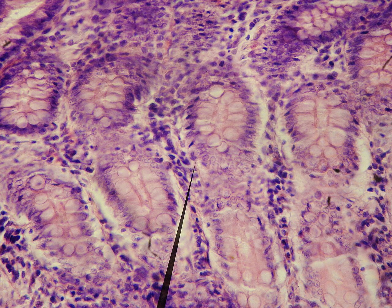
The small intestine has three distinct regions – the duodenum, jejunum, and ileum. The duodenum, the shortest, is where preparation for absorption through small finger-like protrusions called intestinal villi begins. The jejunum is specialized for the absorption through its lining by enterocytes of small nutrient particles which have been previously digested by enzymes in the duodenum. The main function of the ileum is to absorb vitamin B12, bile salts, and whatever products of digestion were not absorbed by the jejunum. The length of the small intestine can vary greatly, from as short as 3 metres (10 feet) to as long as 10.5 m (34+1⁄2 ft), also depending on the measuring technique used. The typical length in a living person is 3–5 m (10–16+1⁄2 ft). The length depends both on how tall the person is and how the length is measured. Taller people generally have a longer small intestine and measurements are generally longer after death and when the bowel is empty. The small intestine develops from the midgut of the primitive gut tube. By the fifth week of embryological life, the entire midgut loop begins to grow longer at a very fast rate, forming a U-shaped fold called the primary intestinal loop. The loop grows so fast in length that it outgrows the abdomen and protrudes through the umbilicus. By week 10, the loop retracts back into the abdomen.
Reader's Guide
The small intestine is central to human digestion and nutrition. It is where most chemical digestion takes place, with enzymes from the pancreas and liver entering via the pancreatic duct. Proteins are degraded into small peptides and amino acids; lipids are broken down into fatty acids and glycerol with the help of bile salts; carbohydrates are degraded into simple sugars. The small intestine's enormous surface area, averaging 30 square metres due to folds, villi, and microvilli, allows efficient absorption of these nutrients into the bloodstream. Its three sections—duodenum, jejunum, and ileum—each have specialized roles: the duodenum prepares food for absorption, the jejunum absorbs most nutrients, and the ileum absorbs vitamin B12 and bile salts. The organ's blood supply comes from the celiac trunk and superior mesenteric artery, ensuring oxygen and nutrients reach its tissues. Understanding the small intestine is essential for grasping how the body extracts energy and building blocks from food, and its dysfunction can lead to malnutrition and other disorders.
Did You Know?
- The small intestine is about 6.5 metres (21 feet) long but is called 'small' because it is narrower in diameter than the large intestine.
- The small intestine has three distinct regions: the duodenum, jejunum, and ileum.
- The small intestine develops from the midgut of the primitive gut tube and rotates anticlockwise during embryonic development.
The Three-Part Architecture
The small intestine is organized into three distinct segments, each with a specialized role. The duodenum, the shortest at roughly 20 to 25 centimeters, takes on a C-shaped form that cradles the head of the pancreas. It is here that gastric chyme first meets pancreatic enzymes and bile, while Brunner's glands secrete an alkaline, mucus-rich fluid to neutralize the acidity of stomach contents. The jejunum stretches about 2.5 meters and serves as the primary site where sugars, amino acids, and fatty acids cross the intestinal lining into the bloodstream, aided by circular folds and finger-like villi that dramatically expand the absorptive surface. The ileum, the longest segment at approximately 3 meters, completes the absorption process by capturing vitamin B12, bile salts, and any residual nutrients the jejunum missed before handing off to the cecum at the ileocecal junction. Together, these three regions span roughly 6.5 meters in a typical adult, folding repeatedly to fit within the abdominal cavity.
The Chemistry of Digestion
The small intestine is the central stage where chemical digestion reaches its peak. Food passes from the stomach through the pyloric sphincter into the duodenum, where it encounters a coordinated wave of digestive secretions. Pancreatic enzymes, released in response to the hormone cholecystokinin, break down proteins, while bile from the liver emulsifies fats into tiny micelles. Another hormone, secretin, generated within the small intestine itself, prompts the pancreas to release additional bicarbonate to further buffer the acidic chyme. The jejunum then takes over as the principal absorption zone, where enterocytes in the mucosal lining pull digested sugars, amino acids, and fatty acids into the bloodstream. The ileum acts as a final sweep, harvesting vitamin B12, bile salts, and any remaining products of digestion. This sequential, hormone-driven choreography ensures that nearly every usable nutrient is captured before waste material moves on to the large intestine.
Embryonic Origins and the Intestinal Loop
The small intestine's remarkable length and coiled arrangement are the legacy of a dramatic embryonic journey. Originating from the midgut of the primitive gut tube, the developing intestine begins its rapid elongation by the fifth week of gestation. The ileum grows so quickly that it forms a U-shaped structure known as the primary intestinal loop, which outpaces the growing abdomen and temporarily protrudes through the umbilicus. By the tenth week, the loop retracts back into the abdominal cavity. During weeks six through ten, the entire structure rotates anticlockwise when viewed from the front of the embryo, and after re-entering the abdomen it completes an additional 180-degree turn. This sequence of growth, protrusion, retraction, and rotation is what produces the characteristic twisted configuration of the adult bowel. The process also explains why the small intestine, despite being roughly 6.5 meters long, must fold many times to occupy the relatively compact space of the abdomen.
Vascular Network and Molecular Identity
The small intestine is sustained by a richly branched vascular system drawn from two major aortic branches: the celiac trunk and the superior mesenteric artery. The duodenum receives dual supply through the superior and inferior pancreaticoduodenal arteries, whose anterior and posterior branches converge and anastomose along the midline. The jejunum and ileum are nourished by branches of the superior mesenteric artery that form layered arches called arterial arcades within the mesentery, from which straight vessels known as vasa recta reach the intestinal wall. At the molecular level, the duodenum expresses roughly 70 percent of the approximately 20,000 protein-coding genes active in human cells, with about 300 genes showing duodenal-specific expression. Proteins such as fatty acid binding protein FABP6 and DEFA6, found in Paneth cell secretory granules, exemplify the specialized molecular toolkit that distinguishes different regions of this remarkably versatile organ.
Gallery
Frequently Asked Questions
Who is Small intestine in Physiology & Medicine?
Small intestine (also called the small bowel) is the long tubular organ nestled between the stomach and the large intestine in the gastrointestinal tract. It is the primary site where digested food is broken down and its nutrients are absorbed into the bloodstream.
What are Small intestine's powers/role?
Its core function is absorbing the vast majority of nutrients from the food we eat. It receives bile and pancreatic secretions through the major duodenal papilla to help finish the digestive process before passing the remaining material onward.
How does Small intestine's story end?
Its narrative arc concludes at the ileocecal valve, where the ileum—its final segment—hands off the leftover chyme to the large intestine. From that junction, the small intestine's active role in nutrient absorption is complete.
Why is Small intestine important to the Physiology & Medicine canon?
Despite its modest diameter of roughly 2.5–3 cm in adults, it stretches 3 to 5 metres in a living person, giving it an enormous folded surface area for absorption. Without it, the body could not efficiently extract the vitamins, minerals, and macronutrients it needs to survive.
What are Small intestine's three parts?
The organ is divided into the duodenum, jejunum, and ileum, each with slightly different structural and functional specializations. Blood supply is delivered by the celiac trunk and the superior mesenteric artery, keeping all three segments well perfused.
More in Physiology & Medicine 1-19
Spotted an error? Know more?
This is a living reference — every entry is fact-audited, and reader corrections feed straight into our audit queue. Suggest an edit · See this site's audit record